Peptide profiler
Quick adme quote
Connect with us


Cellular screening is a critical step in integrated drug discovery, bridging the gap between early biochemical and biophysical screening and more complex phenotypic and translational assays used during lead optimization and candidate selection. By evaluating compounds and biologics in a relevant cellular context, our cellular screening services help you understand not just whether a molecule binds and has a phenotypic response, but also how it behaves in a complex biological system.
Designing and delivering robust, scalable cell-based screening assays that generate biologically meaningful data is our specialty, ensuring you can progress the right compounds faster, reducing late-stage attrition and de-risking downstream development, saving you time and money.
Drug discovery teams often face common challenges when moving from biochemical or biophysical assays into cell-based systems:
By addressing these challenges, providing orthogonal, decision-enabling data that supports confident hit confirmation and lead optimization, the cell-based screening assays that we offer ensure your project is a success.
Cell-based assays are used to assess how compounds influence key biological parameters, including:
These assays capture the impact of cell permeability, metabolism and off-target activity, delivering a more realistic view of compound behavior earlier in the discovery pipeline.
Using advanced imaging, automation and multi-modal readouts, our cellular assays generate high-content phenotypic data that helps you prioritize the most promising leads efficiently and accurately.
Our cellular screening capabilities support secondary screening, hit confirmation and hit-to-lead optimization in preclinical drug discovery, and include:
These assays are both target and modality agnostic, making them applicable across small molecules, biologics and emerging therapeutic approaches.
Learn more about our cell-based screening assays
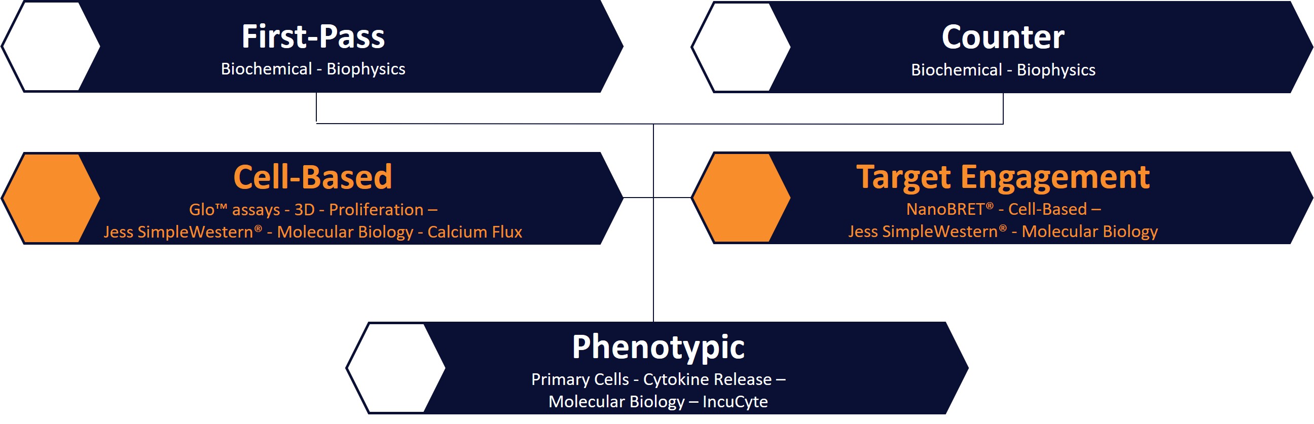
Concept Life Sciences’ assay development and screening group supports the full span of preclinical drug discovery, from hit identification through to lead optimization. Cell-based and target engagement assays form a vital link in this pipeline by enabling:
All assays are supported by streamlined data analysis and recording via a robust ELN/LIMS infrastructure, ensuring seamless data flow between Biology, Chemistry and ADMET teams.
This tightly integrated approach accelerates Design–Make–Test–Analyze (DMTA) cycles and enables faster, better-informed decision-making.
Our scientists understand the nuances that determine cellular assay success including cell density, incubation times, vehicle tolerance, media composition and on-target versus off-target toxicity. This expertise allows us to rapidly establish fit-for-purpose, reproducible assays fully aligned to your project goals.
Cell-based and target engagement assay platforms include, but are not limited to:
By bringing all screening capabilities together at one site, and co-locating Biology with Medicinal Chemistry and ADMET, we help you:
Designed around your program, your timelines and your decision points, our cellular screening services provide high-quality, biologically relevant data you need to move forward with confidence.
Talk to our scientists to discuss how our cellular screening capabilities can accelerate your integrated drug discovery program.
A: Cellular screening evaluates how compounds interact with living cells, providing physiologically relevant data that helps predict efficacy, toxicity, and mechanism of action earlier in drug discovery.
A: By combining advanced imaging, automation, and integration with chemistry and ADMET teams, we reduce DMTA cycles and deliver reliable phenotypic and target engagement data faster.
A: We provide reporter gene assays, target engagement studies (e.g., NanoBRET®), phenotypic screening, cytotoxicity, and cell viability assays — all designed for high reproducibility and translational relevance.
A: Our assays cover immortalized and primary cell lines, GPCRs, ion channels, nuclear receptors, and protein–protein interactions, with read-outs including luminescence, fluorescence, FRET/BRET, calcium flux, and mechanistic studies.