Peptide profiler
Quick adme quote
Connect with us


Concept Life Sciences offers advanced 3D and ex vivo tissue model-based assays that move beyond traditional 2D in vitro systems, delivering more physiologically relevant insights to accelerate your drug discovery process.
Our expert team collaborates closely with you to design assays tailored to your scientific, technical, and strategic goals. By creating tailored 3D and ex vivo model-based assays we help you validate targets and profile lead compounds with confidence to accelerate candidate nomination and provide translational data that informs clinical decision-making, driving success.
Confirming efficacy in complex disease-relevant model systems before advancing to expensive in vivo studies can be complex, time-consuming, and resource-intensive.
Watch the on-demand webinar - 3D Co-Cultures: Choosing the Best Systems & Analysis for Immuno-Oncology - where Darryl Turner, PhD in Veterinary Parasitology and Immunology, Principal Scientist at Concept Life Sciences, discusses the approaches currently employed at Concept Life Sciences, with a particular focus on how we assess different cell types within co-cultures. Emphasis is placed on the inclusion of fibroblasts and immune cells, and how their interaction can influence outcomes.
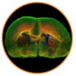
When testing molecules in complex systems, human and rodent models provide powerful insight, especially for CNS-targeted compounds. Organotypic brain slices are the only preclinical model that preserves native brain tissue architecture and diverse cell populations.
Applications include:
Learn more about disease modeling
Contact us for expert guidance
A: Advanced in vitro and ex vivo systems that mimic human tissue architecture, providing more predictive, translational preclinical data.
A: 3D spheroids recreate cellular interactions, tissue gradients, and microenvironments, making preclinical predictions more accurate than 2D cultures.
A: CNS-targeted drug testing, neuroinflammatory studies, toxicity assessment, neuroprotection, and myelination studies